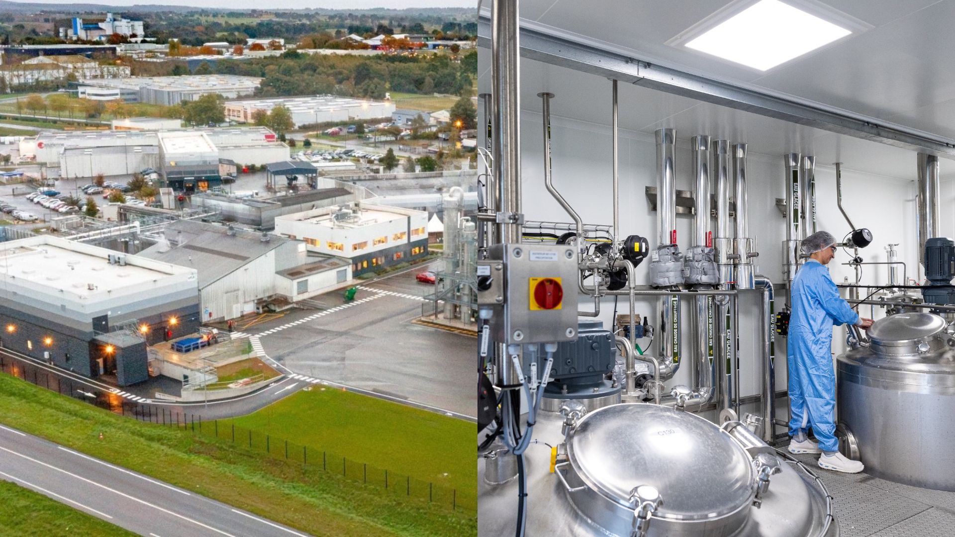
HTL Biotechnology launches HTL8
A new production unit dedicated to pharmaceutical-grade Polynucleotides (DNA) at our Javené site.

Introduction
Glaucoma is the leading cause of blindness throughout the world, after cataract. This is a complex eye condition characterized by an elevated intraocular pressure (IOP), damaging the optic nerves. There are different types of glaucoma, defined by the anatomy of the eye anterior chamber angle’s (open or close) and the major etiology (primary or secondary). Primary open angle glaucoma (POAG) is the most common type, affecting 74% of patients with glaucoma.
Current treatments cannot cure optic nerve damage. However, they can control disease progression by lowering the IOP (the only manageable risk factor) through medication, laser treatment or incisional glaucoma surgeries, such as trabeculectomy. Pharmacotherapy can have undesirable adverse effects, while laser and incisional surgeries involve greater procedural risks and frequent treatment failures. For example, the reoperation rate for trabeculectomy after five years is around 15%. This is why minimally invasive glaucoma surgeries (MIGS) have been developed. MIGS offer a better safety profile, reducing IOP while protecting the eye’s conjunctiva. Canaloplasty, in particular, is a MIGS procedure that uses hyaluronic acid based ophthalmic viscosurgical devices (OVD). [1,2]
The benefits of minimally invasive glaucoma surgeries compared to trabeculectomy
Trabeculectomy used to be the gold standard surgical procedure for glaucoma, associated with a significant reduction in IOP. It involves creating an induced surgical fistula to help aqueous humor drainage. Despite its effectiveness, it can lead to hypotony, choroidal effusion, infection-related bleb formation and endophthalmitis. Patients undergoing trabeculectomy also require more glaucoma medications postoperatively.
Although trabeculectomy remains superior in terms of IOP reduction, MIGS provides a less invasive solution, reducing the recuperation period and the rate of postoperative issues, while also making conjunctival tissue available for use in future surgeries. Another important outcome is that patients require less medication after surgery, which reduces their medication burden and improves their quality of life.
MIGS involves different strategies such as:
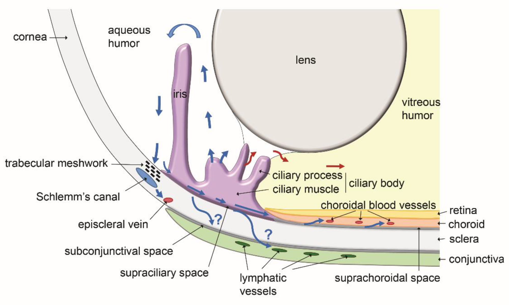
Canaloplasty, for example, is a surgical procedure that involves dilating Schlemm’s canal to improve aqueous humor flow. [3,4]
Canaloplasty and hyaluronic acid based ophthalmic viscosurgical devices
Canaloplasty can be performed using two different approaches : the ab externo (ABeC) technique, which involves external instrumentation and sometimes dissection, and the ab interno (ABiC) technique, which is performed through a small clear corneal incision.
The ab interno approach reduces the risk of complications such as hypotony, infection and postoperative scarring. It can also be combined with cataract surgery, since this procedure is known to reduce IOP in some glaucoma patients. Moreover, due to its relative simplicity, the ABiC technique is considered one of the better options for canaloplasty treatment. The procedure consists of inserting a microcatheter into Schlemm’s canal and then circumnavigating the entire 360 degrees of Schlemm’s canal with the microcatheter before slowly withdrawing it. During removal, aliquots of high-molecular-weight hyaluronic acid-based OVD are delivered into Schlemm’s canal to preserve dilation. [5,6]
Hyaluronic acid is found naturally in the tear film, the outer cornea and the vitreous humor. Its biological compatibility, viscoelasticity, excellent water-binding and wetting properties make it particularly relevant for OVDs. Additionally, the viscosity of the OVD can be adapted to suit the needs of the surgery by adjusting the molecular weight of the biopolymers. Hyaluronic acid is also involved in intercellular interactions, cell-matrix adhesion and cell mobility, paving the way for innovative applications of hyaluronic acid in glaucoma treatment. [7,8]
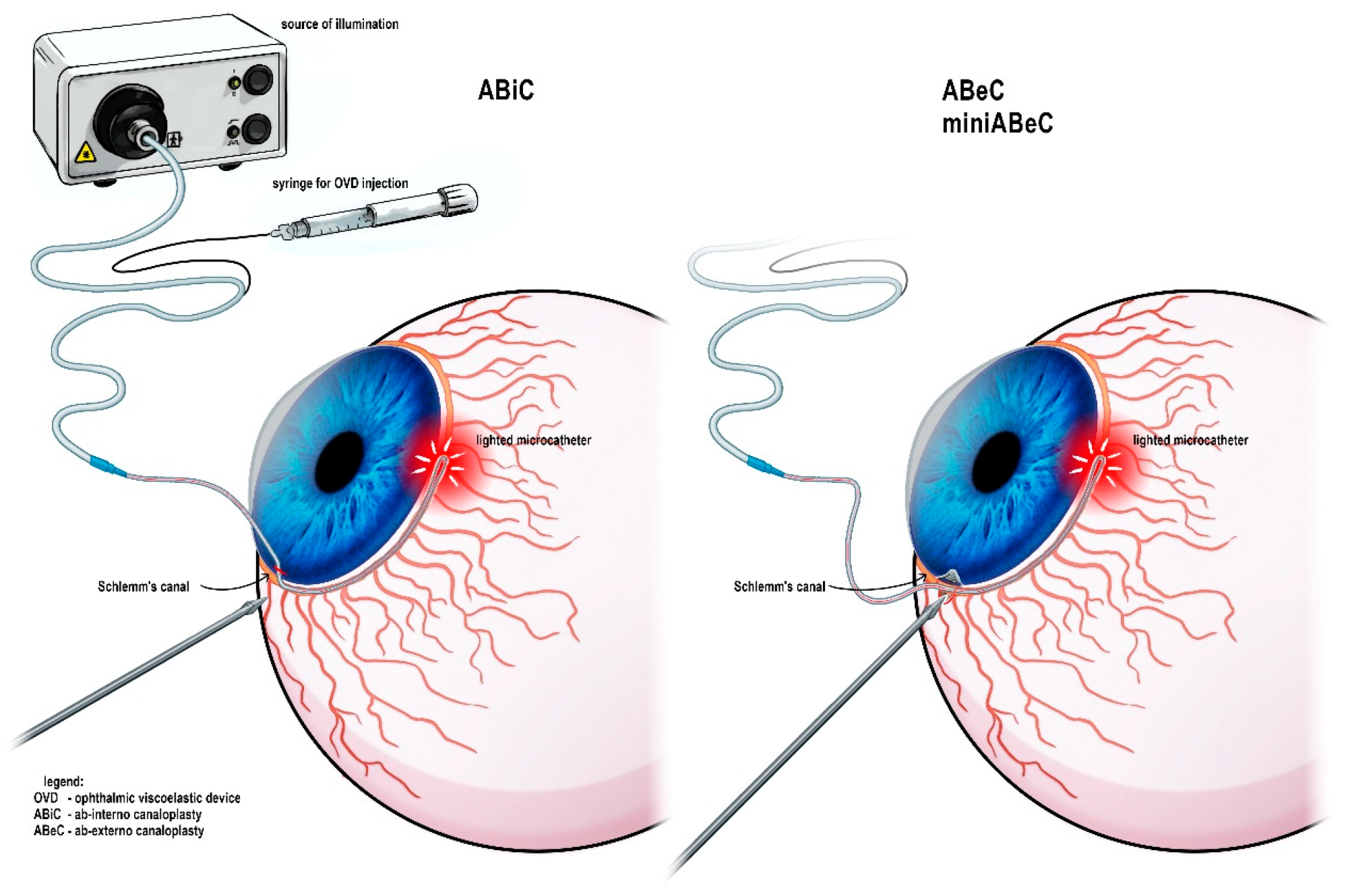
Figure 2 : Instrumentation used during the canaloplasty procedure and variable possible access to the Schlemm’s canal.
Perspectives on glaucoma treatments with hyaluronic acid
The management of glaucoma relies greatly on the topical delivery of medication via eye drops. However, as previously mentioned, pharmacotherapy can have undesirable side effects. Firstly, the residence time of the administered pharmaceutical agent is reduced due to the rapid turnover of the tear film. To improve solubility, retention time and stability, many formulations incorporate additives such as preservatives and surfactants. These additives can cause adverse effects such as irritation, dryness and allergic reactions. In addition, ocular surface disease is highly prevalent among glaucoma patients.
Very high molecular weight hyaluronic acid is a promising alternative approach. It can act as an advanced drug delivery vehicle, increasing the residence time of drugs on the ocular surface thanks to its viscoelastic and mucoadhesive properties. At the same time, the biopolymer provides intrinsic benefits, such as anti-inflammatory activity, protection of the ocular surface, and support for recovery of the corneal nerves. Thus, it has the potential to enable safer and more effective glaucoma therapy while ensuring patient comfort. [9]
Conclusion
MIGS is a revolutionary strategy for reducing IOP while minimizing surgical risk and the patient’s medication burden. Although trabeculectomy remains the preferred option for advanced cases requiring maximum IOP control, MIGS should be considered as the primary surgical option for patients with early-to-moderate glaucoma, comorbidities, or high surgical risk. Canaloplasty is a MIGS procedure that has been shown to be effective and safe in the long term, making it a good choice for most patients with open-angle glaucoma. Moreover, it uses OVDs containing hyaluronic acid, a biopolymer with interesting viscoelastic and biocompatible properties. Hyaluronic acid is also a promising material for developing drug delivery systems for glaucoma treatment, as it enhances residence time and protects ocular tissue.
References:
[1] Wagner, Isabella V., and al. « Updates on the Diagnosis and Management of Glaucoma ». Mayo Clinic Proceedings: Innovations, Quality & Outcomes, vol. 6, nᵒ 6, December 2022, p. 618‑35. DOI.org (Crossref), https://doi.org/10.1016/j.mayocpiqo.2022.09.007.
[2] Dietze J, Blair K, Zeppieri M, and al. Glaucoma. [Updated 2024 Mar 16]. In: StatPearls [Internet]. Treasure Island (FL): StatPearls Publishing; 2024 Jan-. https://www.ncbi.nlm.nih.gov/books/NBK538217/
[3] Chen, Kai-Yang, and al. « Efficacy and Safety of Trabeculectomy versus Micro-Invasive Glaucoma Procedures (MIGPs) in Managing Glaucoma: A Systematic Review and Meta-Analysis ». European Journal of Medical Research, vol. 31, no 1, February 2026, p. 426. DOI.org (Crossref), https://doi.org/10.1186/s40001-026-03868-1.
[4] Gallo Afflitto, Gabriele, and Swarup S. Swaminathan. « Minimally Invasive Glaucoma Surgery ». International Ophthalmology Clinics, vol. 63, no 4, October 2023, p. 33‑60. DOI.org (Crossref), https://doi.org/10.1097/IIO.0000000000000479.
[5] Gallardo, Mark J. « 24-Month Efficacy of Viscodilation of Schlemm’s Canal and the Distal Outflow System with iTrack Ab-Interno Canaloplasty for the Treatment of Primary Open-Angle Glaucoma ». Clinical Ophthalmology, Volume 15, April 2021, p. 1591‑99. DOI.org (Crossref), https://doi.org/10.2147/OPTH.S272506.
[6] Dorairaj, Syril, and al. « A Review of Canaloplasty in the Treatment and Management of Glaucoma ». Journal of Current Glaucoma Practice, vol. 18, no 2, July 2024, p. 79‑85. DOI.org (Crossref), https://doi.org/10.5005/jp-journals-10078-1442.
[7] Hynnekleiv, Leif, and al. « Hyaluronic Acid in the Treatment of Dry Eye Disease ». Acta Ophthalmologica, vol. 100, no 8, December 2022, p. 844‑60. DOI.org (Crossref), https://doi.org/10.1111/aos.15159.
[8] Borkenstein, Andreas F., and al. « Ophthalmic Viscosurgical Devices (OVDs) in Challenging Cases: A Review ». Ophthalmology and Therapy, vol. 10, no 4, December 2021, p. 831‑43. DOI.org (Crossref), https://doi.org/10.1007/s40123-021-00403-9.
[9] Pujol-Martí, Jesús, and Wolfgang G. K. Müller-Lierheim. « Very High Molecular Weight Hyaluronic Acid as an Enhanced Vehicle in Therapeutic Eye Drops: Application in a Novel Latanoprost Formulation for Glaucoma ». Bioengineering, vol. 12, no 9, August 2025, p. 907. DOI.org (Crossref), https://doi.org/10.3390/bioengineering12090907.
Images:
[Figure 1] Subileau, Mariela, et Daniel Vittet. « Lymphatics in Eye Fluid Homeostasis: Minor Contributors or Significant Actors? » Biology, vol. 10, no 7, juin 2021, p. 582. DOI.org (Crossref), https://doi.org/10.3390/biology10070582.
[Figure 2] Kicińska, Aleksandra K., et Marek Rękas. « Safety and Efficacy of Three Modifications of Canaloplasty to Treat Open-Angle Glaucoma: 3-Year Outcomes ». Journal of Clinical Medicine, vol. 12, no 20, octobre 2023, p. 6475. DOI.org (Crossref), https://doi.org/10.3390/jcm12206475.


Glaucoma is currently one of the leading causes of irreversible vision loss worldwide. This chronic disease is characterized by progressive damage to the optic nerve, with elevated intraocular pressure being the main modifiable risk factor for disease development and progression. Management relies on the daily administration of pressure-lowering treatments in the form of eye drops to slow disease progression. Although these treatments have demonstrated their efficacy, their long-term use remains challenged by a major issue: treatment adherence, due in part to the physiological limitations of conventional ocular drug administration.
In response to these limitations, the development of new ophthalmic drug delivery technologies aims to improve drug residence time in the eye and ensure a more consistent release of the active pharmaceutical ingredient. Among these innovations, ophthalmic inserts are emerging as a promising technology. [1]
Skin quality has become one of the main focuses of modern aesthetic medicine. While traditional treatments have often aimed to correct visible signs of ageing, today’s regenerative approaches seek to improve the skin itself by supporting hydration, structure and tissue regeneration.

Hyaluronic acid (HA) has become the dominant biomaterial in injectable soft tissue augmentation, with millions of treatments performed annually worldwide. As a naturally occurring glycosaminoglycan composed of repeating disaccharide units of D-glucuronic acid and N-acetyl-D-glucosamine, native HA possesses exceptional biocompatibility, hygroscopicity, and viscoelastic properties. However, in its unmodified state, HA is rapidly degraded in vivo by endogenous hyaluronidases and reactive oxygen species, with a tissue half-life measured in hours to days. [1,2]
To overcome this limitation and create materials suitable for lasting soft tissue correction, HA must be chemically cross-linked. This process transforms a fluid polysaccharide solution into a structured hydrogel with tuneable mechanical and biological properties. The chemistry of cross-linking is, in many respects, the defining step that determines how a dermal filler will perform in the hands of a clinician and in the tissues of a patient. [2,3]