EcoVadis Platinum Level Achieved in 2026
We have renewed our EcoVadis assessment and achieved the Platinum level!

Introduction :
Dry Eye Disease (DED) is a chronic, multifactorial disorder of the ocular surface that represents a clinical and socioeconomic burden worldwide. Defined by the TFOS DEWS II (Tear Film & Ocular Surface Society Dry Eye WorkShop II) report as a loss of tear film homeostasis, DED is associated with tear film instability, hyperosmolarity, ocular surface inflammation, and neurosensory abnormalities. These interconnected mechanisms contribute not only to patient discomfort but also to progressive damage of the corneal epithelium and visual impairment. [1]
Current data show that up to half of adults report symptoms of dry eye, with an estimated 350 million individuals affected worldwide. In Europe alone, approximately 20% of the population has been diagnosed, highlighting the scale of the condition. Despite available treatments, 25% of patients feel that their management is insufficient, reflecting an important unmet medical need. Moreover, 50% of patients experience daily symptoms, underlining the chronic and persistent nature of DED. [2]
Pathophysiology of DED :
The pathophysiology of DED is complex and involves a wide range of intrinsic and extrinsic factors :
all contribute to disruption of the tear film.
Clinically, the disease is commonly classified into aqueous-deficient and evaporative forms, although a mixed etiology is frequently observed. Regardless of the initial trigger, these conditions converge toward a common pathological pathway characterized by tear film instability and chronic inflammation. [1,2,3]
At the core of DED lies a self-perpetuating inflammatory cycle involving both innate and adaptive immune responses. Tear film disruption and increased osmolarity initiate the release of pro-inflammatory mediators at the ocular surface. This leads to activation and proliferation of T-cells within lymphoid tissues, followed by their migration to the ocular surface, where they sustain inflammation and tissue damage. This vicious cycle progressively impairs epithelial integrity and reduces the capacity of the ocular surface to maintain homeostasis, making disease management particularly challenging.[4]
Therapeutic solutions :
Current therapeutic strategies aim to break this cycle through a stepwise approach tailored to disease severity. [2,3]
HA in the heart of DED :
Within this therapeutic landscape, hyaluronic acid (HA) has emerged as a key component in the management of DED. Naturally present in the tear film and ocular tissues, HA is a high molecular weight glycosaminoglycan characterized by a highly hydrophilic structure. This enables it to retain large amounts of water, thereby improving hydration and contributing to tear film stability. In addition, its non-Newtonian viscoelastic properties allow it to adapt dynamically to blinking: it exhibits low viscosity under shear stress, facilitating uniform distribution across the ocular surface, and higher viscosity at rest, prolonging its residence time. [5]
Beyond its physicochemical properties, HA exerts biological effects. Its ability to interact with cell surface receptors such as CD44 promotes epithelial cell migration and supports corneal wound healing. Furthermore, its mucoadhesive properties enhance adherence to the ocular surface, improving tear film retention and forming a protective barrier against environmental and mechanical stress. By reducing friction during blinking and limiting epithelial microtrauma, HA indirectly contributes to the reduction of inflammatory processes. In addition, by stabilizing the tear film and reducing hyperosmolarity, it helps attenuate the activation of inflammatory pathways, thereby supporting restoration of ocular surface homeostasis.[3,5,6]
Clinical evidence consistently supports the efficacy and safety of HA-based formulations in DED. These treatments have been shown to improve tear film break-up time and corneal staining, and subsequently decreases dryness, irritation, and visual discomfort. As such, HA is no longer considered merely a lubricant but rather a multifunctional therapeutic agent addressing several key mechanisms of the disease.[6,7]
Conclusion :
To put it in a nutschell Dry Eye Disease is a complex condition requiring a comprehensive and multifactorial treatment approach. Hyaluronic acid plays a central role within this paradigm by combining hydration, protection, and biological activity. Its ability to act simultaneously on tear film stability, epithelial repair, and inflammation makes it a cornerstone of current and emerging therapeutic strategies aimed at restoring ocular surface health.
Bibliography :
[1] Acuité. Sécheresse oculaire : le mal silencieux qui touche un adulte sur deux.
[2] LeMedecin.fr. Syndrome de l’Œil Sec : Symptômes, Traitements et Innovations 2025.
[3] Sheppard J, Lee BS, Periman LM. Dry eye disease: identification and therapeutic strategies for primary care clinicians and clinical specialists. Ann Med. 2023.
[4] Periman LM, Perez VL, Saban DR, Lin MC, Neri P. The Immunological Basis of Dry Eye Disease and Current Topical Treatment Options. J Ocul Pharmacol Ther. 2020.
[5] Utheim TP et al. Hyaluronic acid in the treatment of dry eye disease. Acta Ophthalmol. 2022;100:844–860.
[6] Kim DJ et al. Development of a novel hyaluronic acid membrane for the treatment of ocular surface diseases. Sci Rep. 2021.
[7] Zhang Y et al. Different concentrations of hyaluronic acid eye drops for dry eye syndrome: a systematic review and meta-analysis. J Ophthalmol. 2023

Hyaluronic acid (HA) has become the dominant biomaterial in injectable soft tissue augmentation, with millions of treatments performed annually worldwide. As a naturally occurring glycosaminoglycan composed of repeating disaccharide units of D-glucuronic acid and N-acetyl-D-glucosamine, native HA possesses exceptional biocompatibility, hygroscopicity, and viscoelastic properties. However, in its unmodified state, HA is rapidly degraded in vivo by endogenous hyaluronidases and reactive oxygen species, with a tissue half-life measured in hours to days. [1,2]
To overcome this limitation and create materials suitable for lasting soft tissue correction, HA must be chemically cross-linked. This process transforms a fluid polysaccharide solution into a structured hydrogel with tuneable mechanical and biological properties. The chemistry of cross-linking is, in many respects, the defining step that determines how a dermal filler will perform in the hands of a clinician and in the tissues of a patient. [2,3]
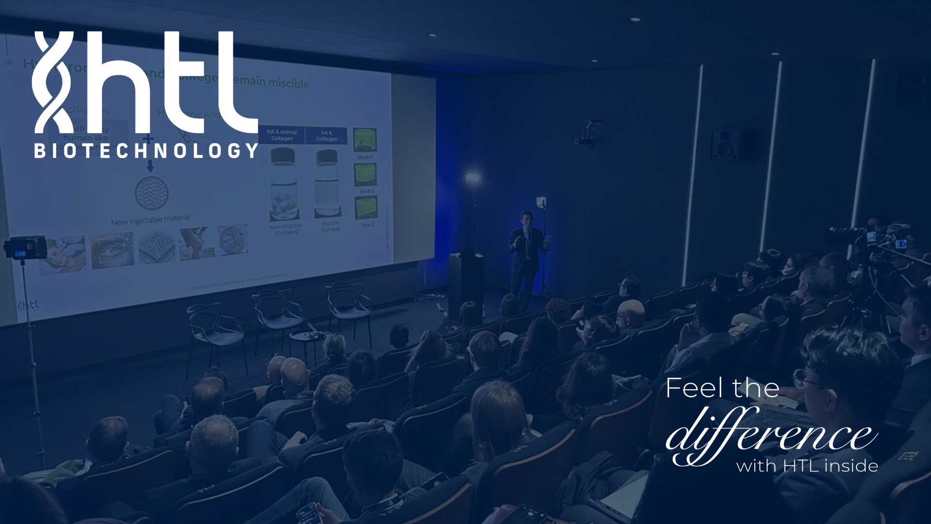
On January 29, 2026, during IMCAS World Congress in Paris, HTL Biotechnology hosted its first scientific session dedicated to the future of regenerative and aesthetic medicine.
Bringing together experts from industry and clinical practice, the session explored how advanced biopolymers are helping shift aesthetic medicine toward a more regenerative approach, focused on skin quality, tissue repair, and long-term biological function.